lined link to go to subject)
Chapter 1. Vision
System Design
Chapter 2. Biological Eye Designs
Chapter
3. Eye
Design
Illustrations
A.
Plant
light sensing
1.
Grass, simple vines,
and stems
2.
Flowers
B.
Lower
animal eyes
1.
Flatworms
2.
Clams and Scallops
3.
Nautilus
4.
Shrimp
5.
Crab
6.
Octopus and
giant squid
7.
Spiders
8.
Scorpions
8.
Brittle Star
C.
Insect
eyes
1.
Bees
2. Dragonflies
3. Butterflies
4.
Flies
5.
Ants
6.
Moths
7.
Beetles
8.
Wasp
D. Fish
eyes
1.
Shark
2.
Flounder
3.
Four-eyed fish
E.
Amphibian
eyes
1. Frog
2.
Salamander
F.
Reptile
eyes
1. Boa
constrictor
2.
Rattle
snake
3.
Lizard
4.
Turtle
5.
Crocodile
and
alligators
G. Bird
eyes
1.
Eagles
2.
Hummingbirds
3. Owls
4.
Ostrich
5.
Cormorants
H.
Mammal
eyes
1.
Whales
2.
Elephants
3. Lions,
tigers, and
other cats
4.
Monkeys
5. Rats
and mice
6. Bats
7.
Tarsier
I.
Human
eyes
1. Iris
2. Lens
3.
Retina
Chapter
4. Eye
Reproduction
Chapter
5. Optical
Systems
Design
Chapter
6. The Eye Designer
Related
Links
Appendix
A - Slide Show & Conference Speech by Curt Deckert
Appendix
B - Conference Speech by Curt Deckert
Appendix
C - Comments From Our Readers
Appendix
D - Panicked Evolutionists: The Stephen Meyer Controversy
Chapter 3
Section I
(Click on PICTURE IN TEXT to bring up LARGE PICTURE)
3. EYE DESIGN ILLUSTRATIONS
I. Human Eyes
Man has one of the most
remarkable vision systems in the world. The human eye's key features
include a highly-corrected optical design, repeatable geometry of
materials, control by the brain, processing of retina information,
interfacing with the brain from six >different levels of sensor
cells in the retina, color vision, compression of data going to
the brain,
and the highly specific material makeup and orientation which enable
each eye to function and memory of scenes to take place. (Fig 3.57a
from p. 292, The Eyes
& Visual Optical Instruments,
Cambridge Press 1997, by George Smith and David Atchison)
(Fig 3.56 from p. 21, Science
and Medicine, J/F 2000, Retinal
Transplantation by R. B. Aramant)

Layers (Same as Figs 1.7b & 3.60b) 
Eye Diagram |
Sensor Cells in the Retina |
|
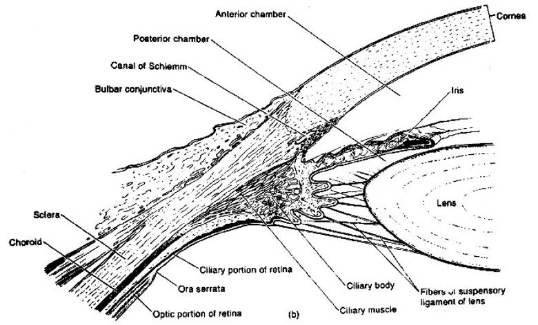
At maturity, adult
human eyeballs are approximately 0.9 inch (24mm) in diameter and
slightly flattened in the front and back. Each of its retina layers is
unique. The outer fibrous layer encasing and protecting the eyeball
consists of the cornea and the sclera. The front one-sixth of the
fibrous layer is the transparent cornea, which functions as a
correction lens to help bend incoming light onto the lens inside the
eye to form a sharp high-resolution image on the retina. Then a fine
membrane covers the cornea. The remaining fibrous layer of
the eye is a dense, tough, opaque coating visible as the white of the
eye. Its outer layer contains blood vessels that produce a "blood-shot
eye" when the eye is irritated. The middle layer of the eyeball is
densely pigmented, well supplied with blood, and includes major complex
structures. The innermost layer includes the retina. Internally, the
eye consists of a front cavity filled with watery aqueous fluid. The
rear cavity is filled with gel-like vitreous fluid. The internal
pressure (the intra-ocular pressure) exerted by the fluid inside the
eye supports the shape of the front cavity, while the fluid with the
holding tissue holds the shape of the rear chamber. An irregular-shaped
eyeball results in ineffective focusing of light onto the retina. One
can be "near sighted" or "far sighted". Both conditions are corrected
with glasses or contacts. These conditions can require spherical and/or
cylindrical corrections. Focusing problems can also come from muscles that control the eye. This condition is also correctable with contacts or glasses. Conditions such as "lazy eye" or "crossed eyes" require special means of correction. A model of the major components of the human eye are further detailed to illustrate the overall vision system in familiar terms. (Fig 3.57b-c adapted from 1999 Eye Poster from Anatomical Chart Co. Skokie, IL) |
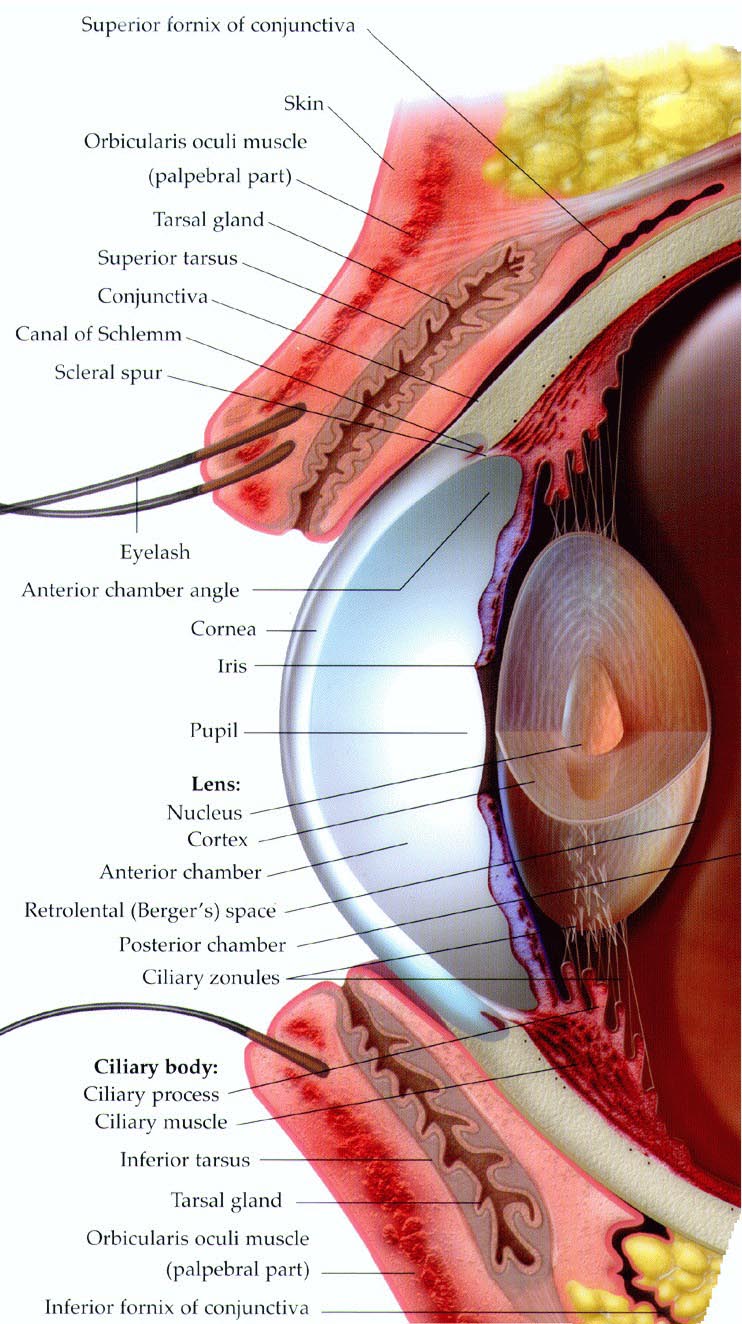
Eye Diagram 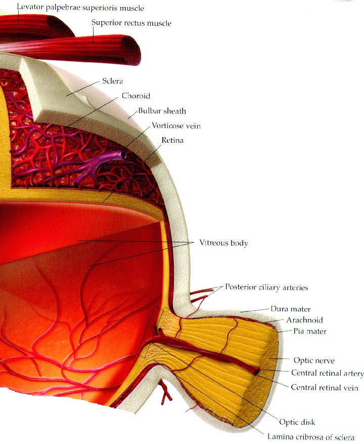
Eye Diagram |
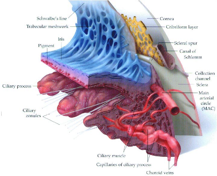
The iris is a circular, adjustable diaphragm with a central aperture (the pupil). It is located in the chamber behind the cornea. The iris gives the eye its color, which depends on the amount of pigment present. If the pigment is dense, the iris is brown. If there is little pigment, the iris is blue. In some cases there is no pigment at all, so the eye is light. Different pigments color eyes in various ways to create the eye colors you see, such as gray, green, etc. In bright light, the iris muscles constrict the pupil, thereby reducing the amount of light entering the eye. Conversely, the pupil enlarges in dim light, to increase the amount of incoming light allowed to go the retina. As light to the retina is reduced, the ability to see color decreases.
The iris is the extension of a large, smooth muscle, which also connects to the lens via a number of suspensor ligaments. These muscles expand and contract to change the shape of the lens, to adjust the focus of images onto the retina. A thin membrane lying beyond the lens provides a light-tight environment inside the eye, thus preventing stray light from confusing or interfering with visual images on the retina. This is extremely important for clear high-contrast vision with good resolution or definition.
The most frontal chamber of the eye, immediately behind the cornea and in front of the iris, contains a clear watery fluid that facilitates good vision. It helps to maintain eye shape, regulating the intra-ocular pressure, providing support for the internal structures, supplying nutrients to the lens and cornea, and disposing of the eye's metabolic waste. The rear chamber of the front cavity lies behind the iris and in front of the lens. It helps provide optical correction for the image on the retina. Some recent optical designs also use coupling fluids for increased efficiency and for better correction. (Fig 3.58a from p. 146, Iridology, Vol. 2, 1982, published by Jensen Enterprises, Escondido, CA 92027) (Fig 3.58b adapted from 1999 Eye Poster from Anatomical Chart Co. Skokie, IL)
Iris Mechanism |
Iris Mechanism |
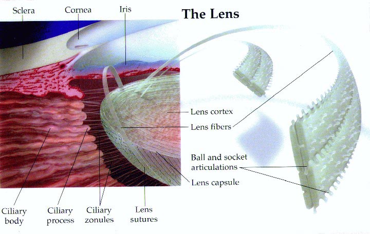
The typical bi-convex (curving outward on both surfaces) lens is a crystal-clear, transparent optical element that is semi-solid and flexible. It is shaped like an elongated sphere. The entire surface of the lens is smooth and shiny, contains no blood vessels, and is encased in an elastic membrane. The lens is held in place by suspensor ligaments that can cause the lens to either fatten or become thin. Complex control systems automatically change its focal length to precisely focus light images on the retina according to where the brain is directing the eye to see. Many variations in human sight due to lens imperfections are now correctable to near perfect vision using new laser techniques, contact lenses, or conventional glasses.
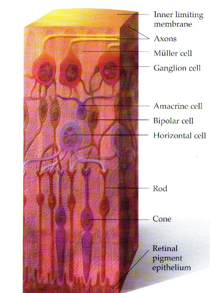
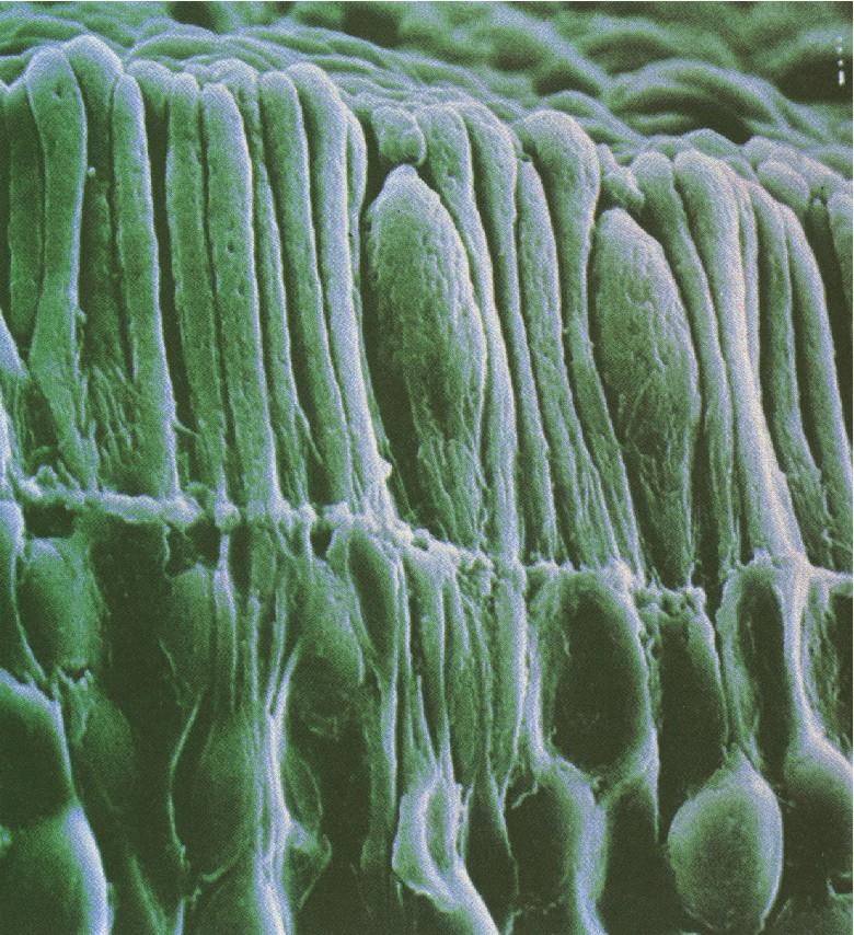
3. Retina
The retina is the
innermost layer making up the eye optical path. It is a thin, delicate,
extremely complex sensory tissue composed of six layers of light
sensitive cells. The retina encircles the rear portion of the eye.
Photoreceptor cells in the rods and cones convert light first to
chemical energy and then electrical energy. Rods function in dim light,
allowing limited night vision. Typically, rods are used to see the
stars; rods do not detect color, but they do detect movements and fine
detail. There are about 126 million rods in each eye and about 6
million cones. This compares to only 1 million sensors in more common
digital cameras. Cones function best in bright light and allow color
vision. Cones are most heavily concentrated in a tiny hollow in the
rear part of the retina.
|
Dense fields of both
rods and cones are found in a circular region surrounding this
high-resolution area. Continuing outward, the cone density decreases
and the ratio of rods to cones increases until both rods and cones
disappear completely at the edges of the retina. This enables us to see
much more detail over a limited field of view than most TV cameras are
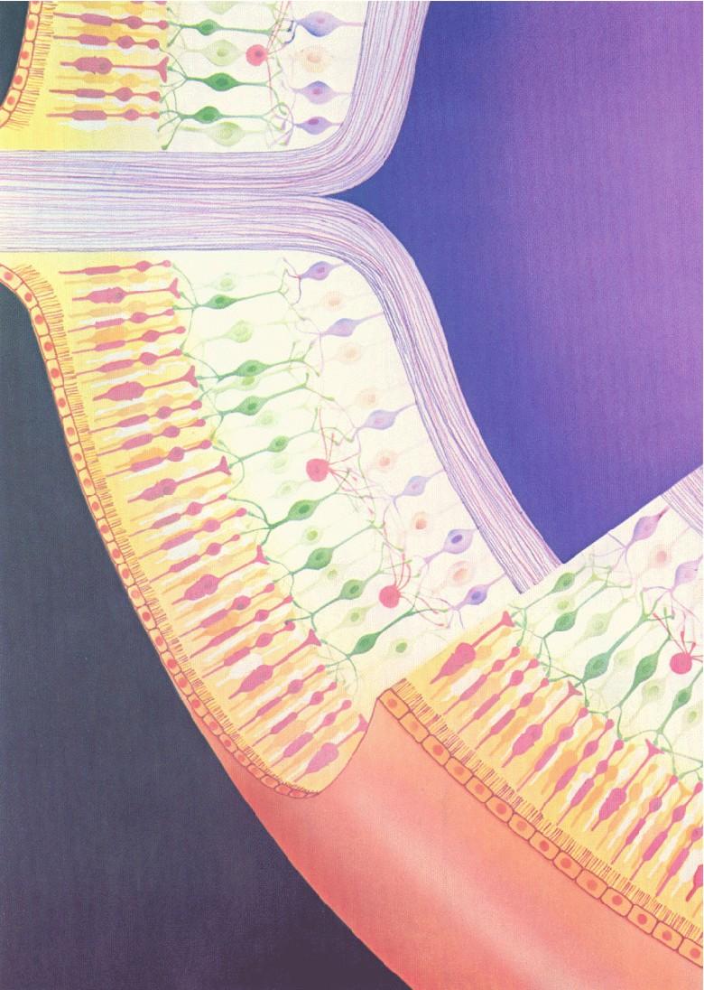
able to resolve. The optic nerve connects the eye to the brain. Thousands of fibers of the optic nerve cells run from the surface of the retina and converge to exit the eye at the optic disc (or blind spot), an area about 0.06 in (1.5mm) in diameter located at the lower rear portion of the retina. The fibers of this nerve are made up of a large number of cells, each having thousands of connections to carry electrical impulses from the retina to the brain. If the optic nerve is severed, vision is permanently lost. The human eye vision system preprocesses the six different levels of sensing in the retina in parallel before information goes to the brain for final processing. These six levels represent six different cell types that make up the retina sensor. Each sensor layer plays a different role in seeing and recognition. Compression of data from each of these layers of sensors results in considerable compression of key visual data going to the brain. This parallel processing allows a rapid means of recognition of complex information. With optical help such as from telescopes, we can further explore our universe. Likewise, we use microscopes to see minute building blocks of eyes such as cells. In comparison with optical instruments, the angular coverage of natural eyes is typically wider than most film and video cameras that are used to record specific events. Our vision systems are an example of irreducible complexity not capable of creation by mutation and natural selection. (Figures 3.59b, 3.60a, and 3.61a from p. 136 and 137, Iridology, Vol. 2, 1982, published by Jensen Enterprises, Escondido, CA 92027) (Fig 3.59a, c and 3.60b adapted from 1999 Eye Poster from Anatomical Chart Co. Skokie, IL) (Figs 3.61b by permission of James T Fulton, Dir of Research Vision Concepts) |
Retina diagram 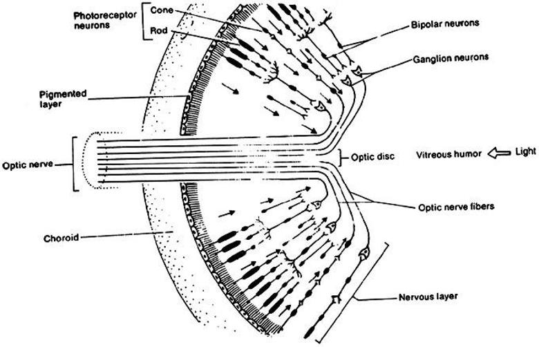
Retina diagram 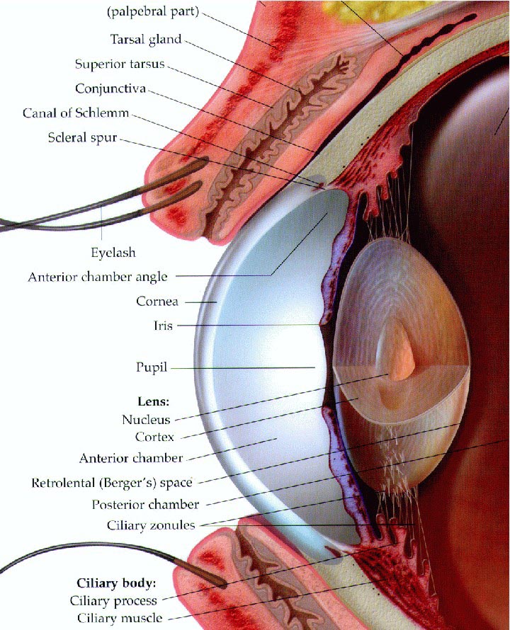
Lens Section 
Sensor Pattern |
Retina diagram for perspective of rods and cones |
Retina Layers (Same as Figs 1.7b & 3.56b) |
Retina Rods and Cones |
Several people have researched image detection and processing technology found in nature with the idea of using it for new system development. Working toward a complete understanding of eyes is certainly a challenge. One article, LIFE LESSONS [click to download PDF file], written by Don Wolpert for the Feb. 2002 issue of OE Magazine, a SPIE publication, has some very interesting ideas. He has done considerable work in this area.
For an intesting article by Peter W. V. Gurney on the retina, go to Is Our ‘Inverted' Retina Really ‘Bad Design'?